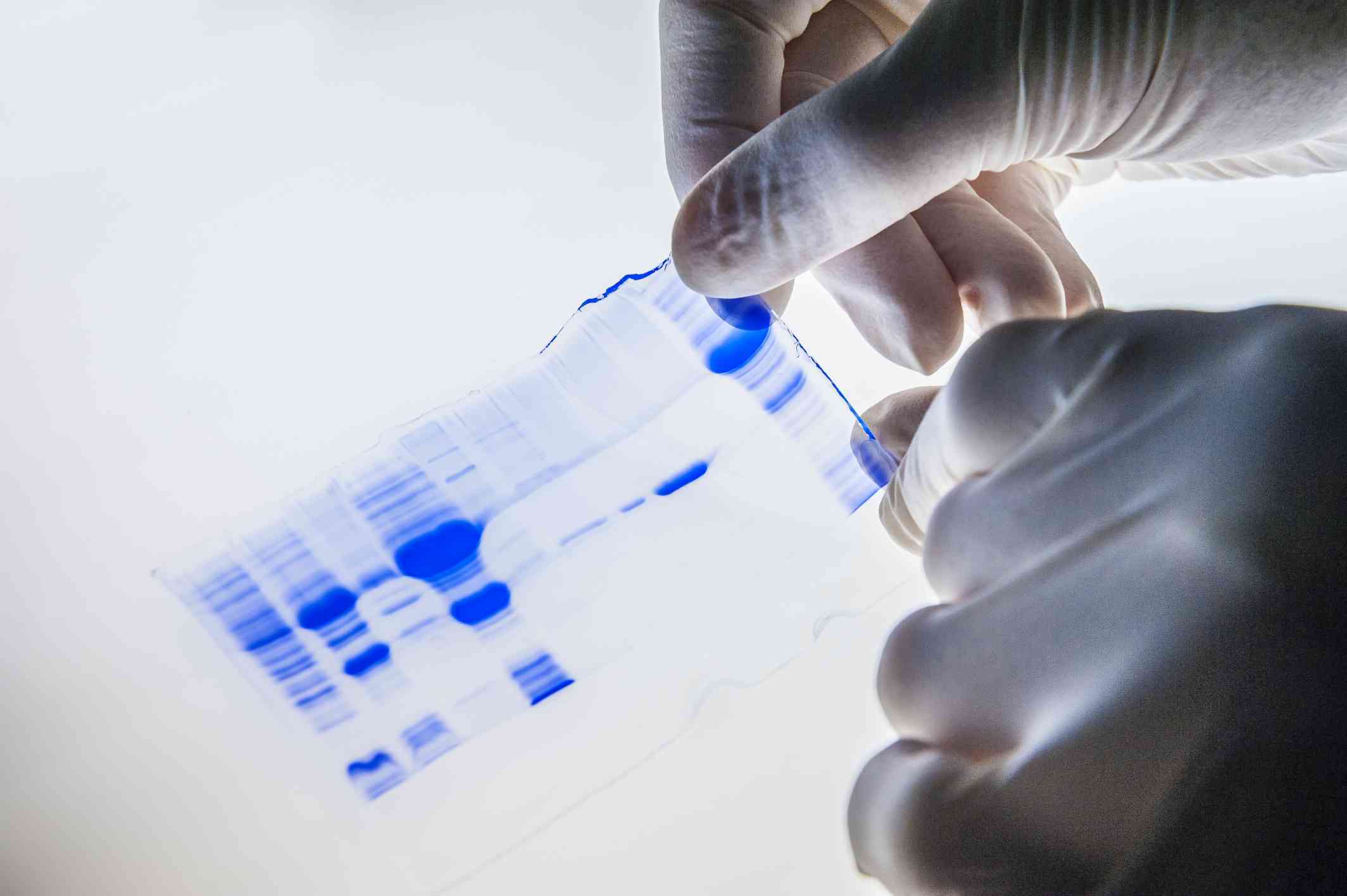
Western blot (WB) is a laboratory technique used by life science researchers and diagnostic laboratories to detect specific proteins within a homogenate or extract of a biological sample. Sample proteins must be extracted from their source using an optimized lysis buffer, separated within a gel using an electrophoresis unit and transferred to a membrane via a “blotting” procedure. Proteins can be separated within a gel by the size of their individual polypeptide subunits to determine their molecular weight (MW), or they can be separated based on their native, tertiary form such that subunit interaction and conformation are preserved. The latter form of separation is determined by their charge-to-mass ratio. After the proteins have been immobilized on the membrane, the proteins of interest can be identified through immunological detection using antibodies conjugated for visualization via chromogenic, chemiluminescent, fluorescent or radioactive mechanisms.

Our WB Resources
| Related Sections | Contents |
| Western Blot Protocol |
Cover the whole process of the experiment:
|
| Western Blot Troubleshooting |
The most common problems and detailed solutions:
|
| Sample Preparation in Western Blot Assay |
The most important part of sample preparation is to fully extract the target protein and avoid protein degradation:
|
| Running an SDS-PAGE Gel in Western Blot Assay |
Factors to consider during electrophoresis:
|
| Protein Transfer from Gel to Membrane in Western Blot Assay |
Transfer of Proteins:
|
| Western Blot Illustrated Assay | Step-by-step instructions |
| Immunological Detection |
Normal immunological detection methods:
|
| Stripping and Reprobing | Numerous advantages of striping and reprobing blots are listed |
| Western Blot Storage | Western blot membrane storage protocol for future analysis or reprobing |
| Quality Control |
|
Considerations Before Starting Your WB Experiment
In the WB assay, it is important to block the unreacted sites on the membrane to reduce the amount of non-specific binding of proteins during subsequent steps in the assay. A variety of blocking buffers ranging from milk or normal serum to highly purified proteins have been used to block unreacted sites on a membrane. The blocking buffer should improve the sensitivity of the assay by reducing background interference. There is no perfect blocking solution suitable for every WB experiment. The proper choice of blocker for a given blot depends on the antigen itself and on the type of enzyme conjugate to be used. For example, with applications using an alkaline phosphatase (AP) conjugate, a blocking buffer in Tris buffered saline (TBS) should be selected because phosphate buffered saline (PBS) interferes with AP. The ideal blocking buffer will bind to all potential sites of non-specific interaction, eliminating background altogether without altering or obscuring the epitope for antibody binding. The most important parameter when selecting a blocker is the signal-to-noise ratio, which is measured as the signal obtained with a sample containing the target analyte as compared to that obtained with a sample without the target analyte.
WB consists of a series of incubations with different immunochemical reagents separated by wash steps. Washing steps are necessary to remove unbound reagents and reduce background, thereby increasing the signal-to-noise ratio. Insufficient washing will allow high background but excessive washing may result in decreased sensitivity caused by elution of the antibody and/or antigen from the blot. Generally, washing is performed in a physiologic buffer such as tris buffered saline (TBS) or phosphate buffered saline (PBS) with detergent such as 0.05% Tween-20 to help remove non-specifically bound material. Another common technique is to use a dilute solution of the blocking buffer along with some added detergent.
Both polyclonal antibody (pAb) and monoclonal antibody (mAb) work well for WB. PAbs are less expensive and less time-consuming to produce and they often have a high affinity for the antigen. MAbs are valued for their specificity, purity and consistency that result in lower background. Crude antibody preparations such as serum or ascites fluid are sometimes used but the impurities present may increase background.
The choice of secondary antibody also depends upon the type of label that is desired. AP and horseradish peroxidase (HRP) are the two enzymes that are used extensively as labels for protein detection. Commercial chromogenic, fluorogenic and chemiluminescent substrates are available for use with either enzyme. More sensitive detection systems require that less antibody be used.