Pax3 Antibodies
Background
Pax3 functions as a transcription factor which drives essential developmental processes in vertebrate embryos. Through its regulatory function Pax3 controls gene expression to develop neural crest cells and muscle tissues which enable proper organogenesis and tissue differentiation. The development of both the nervous system and skeletal muscles depends on Pax3. Mutations in Pax3 are associated with developmental disorders such as Waardenburg syndrome. Scientists discovered Pax3 through molecular cloning in the early 1990s and since then have conducted extensive research which advanced our knowledge of developmental biology and gene regulation and tissue formation and repair mechanisms.
Structure of Pax3
The Pax transcription factor has a weight of, around 48 kDa. Its molecular weight may vary slightly across species due to variations, in amino acid sequences; however the differences are generally not substantial.
| Species | Human | Mouse | Zebrafish | Chicken | Cow |
| Molecular Weight (kDa) | 53 | 53 | 53 | 53 | 53 |
| Primary Structural Differences | Highly conserved across mammals, with minimal sequence variations | Minor amino acid differences compared to human, but retains similar function | Slight functional differences due to evolutionary adaptations, particularly in expression patterns during development | Adapted for avian-specific developmental processes, with unique expression patterns during embryogenesis | Highly similar to human, with minimal sequence variations |
The Pax has sections that serve functions and follows a structured design. It has a paired domain and a homeodomain at the start that help in binding to DNA segments. In the end region of Pax, there is a transactivation domain that controls gene expression. Additionally, there is a region rich in proline, serine, and threonine (PST) that affects its ability to regulate transcription. Pax can team up with itself or other members of the Pax family to increase the affinity for binding to DNA segments. The protein's role is controlled by changes that occur after translation, like phosphorylation and other modifications that affect how stable and active it is. Through interactions with transcription factors and chromatin factors, Pax interacts to coordinate gene activity during growth and illness.
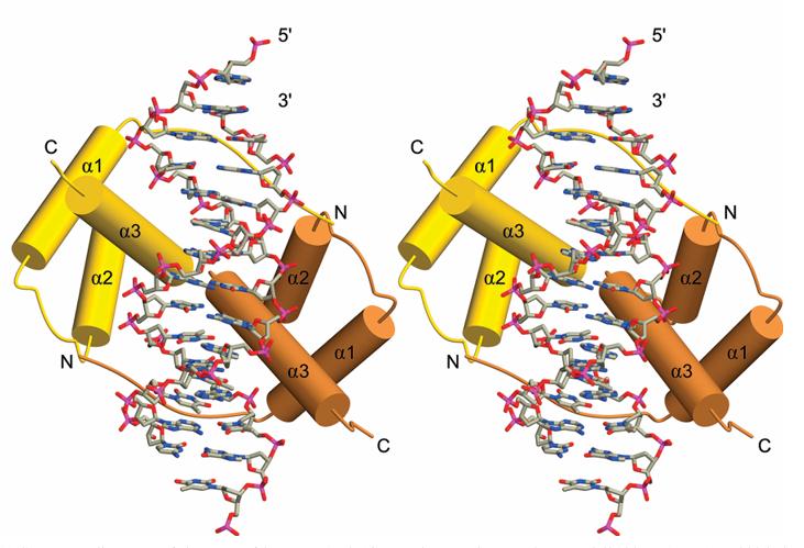 Fig. 1 Stereo view of the PAX3 HD-DNA complex.1
Key structural properties of pax3:
- Features N-terminal transcriptional repression domain.
- Has C-terminal transactivation domain.
- Undergoes phosphorylation, ubiquitination, acetylation.
- Contains paired box and homeodomain for DNA binding.
- Interacts with transcription factors and chromatin factors.
- Forms dimers with Pax3 or Pax7.
Functions of Pax3
The transcription factor Pax3 functions to direct cell differentiation and tissue formation mainly during embryonic development. Pax3 functions as a crucial factor for neural crest cell migration and muscle tissue development while also being linked to cancer development.
| Function | Description |
| Embryonic Development | Regulates gene expression essential for the formation of the neural tube, skeletal muscles, and other tissues during early embryogenesis |
| Cell Differentiation | Controls the specification and differentiation of cell types, particularly in neural crest cells and muscle precursor cells |
| Neural Crest Cell Migration | Guides neural crest cells to their target locations, contributing to the development of the peripheral nervous system and craniofacial structures |
| Muscle Development | Essential for muscle formation, including skeletal muscle development and satellite cell regulation in muscle repair |
| Cancer Development | Aberrant expression or mutations in Pax3 can contribute to tumorigenesis, such as in alveolar rhabdomyosarcoma |
Pax3 shows a dynamic expression pattern during embryogenesis which distinguishes it from other transcription factors because of its essential role in neural crest and muscle development.
Applications of Pax3 and Pax3 Antibody in Literature
1. Kahsay, Abraha, et al. "Pax3 loss of function delays tumour progression in kRAS-induced zebrafish rhabdomyosarcoma models." Scientific Reports 12.1 (2022): 17149. https://doi.org/10.1038/s41598-022-21525-5
The research examines Pax3 function in kRAS-induced rhabdomyosarcoma zebrafish models which shows that tumour progression slows down when Pax3 function is lost thus making it a promising therapeutic target for cancer treatment.
2. Adams, Jason S., Sterling N. Sudweeks, and Michael R. Stark. "Pax3 isoforms in sensory neurogenesis: expression and function in the ophthalmic trigeminal placode." Developmental Dynamics 243.10 (2014): 1249-1261. https://doi.org/10.1002/dvdy.24108
This article delves into the exploration of how Pax3 isoformsre expressed and operate in neurogenesis, within the ophthalmic trigeminal placode region to uncover their significance in the initial stages of neural growth and specialization.
3. Boudjadi, Salah, et al. "The expression and function of PAX3 in development and disease." Gene 666 (2018): 145-157. https://doi.org/10.1016/j.gene.2018.04.087
The article thoroughly explores PAX 23 by discussing where it can be found in the body and its significance, in stages of growth and health conditions related to it demonstrating how important PAX 23s involvement in guiding cells during early development and muscle formation processes truly are, along with its links to specific disorders like Waardenburg syndrome and cancerous growths in muscle tissue thus showcasing its potential as a target for treatment and a tool, for diagnosing illnesses.
4. Udagawa, Tomokatsu, et al. "Pax3 deficiency diminishes melanocytes in the developing mouse cochlea." Research Square (2023): rs-3. https://doi.org/10.21203/rs.3.rs-2990436/v1
This article investigates how Pax3 deficiency reduces melanocyte numbers in the developing mouse cochlea, highlighting its critical role in melanocyte development and cochlear function.
5. Xia, Liang, et al. "PAX3 is overexpressed in human glioblastomas and critically regulates the tumorigenicity of glioma cells." Brain research 1521 (2013): 68-78. https://doi.org/10.1016/j.brainres.2013.05.021
This article reveals that PAX3 is overexpressed in human glioblastomas and plays a critical role in regulating the tumorigenicity of glioma cells, suggesting its potential as a therapeutic target.
Creative Biolabs: Pax3 Antibodies for Research
Creative Biolabs excels in manufacturing superior pax3 antibodies for research and industrial uses. Our range features both monoclonal and polyclonal antibodies, specifically designed for applications in ELISA, Flow Cytometry, Western blot, immunohistochemistry, and various other diagnostic techniques.
- Pax3 Antibody Customization: Tailored solutions to match your unique research goals.
- Mass Production: Industrial-scale antibody production for our partners.
- Technical Guidance: Professional advice for protocol optimization and troubleshooting.
- Aliquoting Services: Thoughtfully-sized aliquots to ensure long-term stability and reliable outcomes.
For additional information regarding our Pax3 antibodies, bespoke services, or technical assistance, please reach out to us via info@creative-biolabs.com.
Reference
- Birrane, Gabriel, Aditi Soni, and John AA Ladias. "Structural basis for DNA recognition by the human PAX3 homeodomain." Biochemistry 48.6 (2009): 1148-1155. https://doi.org/10.1021/bi802052y
Anti-Pax3 antibodies
 Loading...


Hot products 
-
Mouse Anti-BCL6 Recombinant Antibody (CBYY-0442) (CBMAB-0445-YY)

-
Mouse Anti-CDKL5 Recombinant Antibody (CBFYC-1629) (CBMAB-C1689-FY)

-
Mouse Anti-ADIPOR1 Recombinant Antibody (V2-179982) (CBMAB-A1368-YC)

-
Mouse Anti-BACE1 Recombinant Antibody (CBLNB-121) (CBMAB-1180-CN)

-
Rabbit Anti-ENO2 Recombinant Antibody (BA0013) (CBMAB-0272CQ)

-
Mouse Anti-ADGRE2 Recombinant Antibody (V2-261270) (CBMAB-C0813-LY)

-
Mouse Anti-DDC Recombinant Antibody (8E8) (CBMAB-0992-YC)

-
Mouse Anti-C5B-9 Recombinant Antibody (CBFYA-0216) (CBMAB-X0304-FY)

-
Mouse Anti-ADGRL2 Recombinant Antibody (V2-58519) (CBMAB-L0166-YJ)

-
Rat Anti-CD300A Recombinant Antibody (172224) (CBMAB-C0423-LY)

-
Mouse Anti-CD24 Recombinant Antibody (ALB9) (CBMAB-0176CQ)

-
Mouse Anti-DLC1 Recombinant Antibody (D1009) (CBMAB-D1009-YC)

-
Mouse Anti-FTH1 Recombinant Antibody (CBXF-1896) (CBMAB-F3426-CQ)

-
Mouse Anti-BCL6 Recombinant Antibody (CBYY-0435) (CBMAB-0437-YY)

-
Mouse Anti-8-oxoguanine Recombinant Antibody (V2-7719) (CBMAB-1898CQ)

-
Mouse Anti-Acetyl SMC3 (K105/K106) Recombinant Antibody (V2-634053) (CBMAB-AP052LY)

-
Rabbit Anti-BRCA2 Recombinant Antibody (D9S6V) (CBMAB-CP0017-LY)

-
Mouse Anti-DLG1 Monolconal Antibody (4F3) (CBMAB-0225-CN)

-
Rat Anti-FABP3 Recombinant Antibody (CBXF-2299) (CBMAB-F1612-CQ)

-
Mouse Anti-ATP1B1 Recombinant Antibody (E4) (CBMAB-0463-LY)

- AActivation
- AGAgonist
- APApoptosis
- BBlocking
- BABioassay
- BIBioimaging
- CImmunohistochemistry-Frozen Sections
- CIChromatin Immunoprecipitation
- CTCytotoxicity
- CSCostimulation
- DDepletion
- DBDot Blot
- EELISA
- ECELISA(Cap)
- EDELISA(Det)
- ESELISpot
- EMElectron Microscopy
- FFlow Cytometry
- FNFunction Assay
- GSGel Supershift
- IInhibition
- IAEnzyme Immunoassay
- ICImmunocytochemistry
- IDImmunodiffusion
- IEImmunoelectrophoresis
- IFImmunofluorescence
- IGImmunochromatography
- IHImmunohistochemistry
- IMImmunomicroscopy
- IOImmunoassay
- IPImmunoprecipitation
- ISIntracellular Staining for Flow Cytometry
- LALuminex Assay
- LFLateral Flow Immunoassay
- MMicroarray
- MCMass Cytometry/CyTOF
- MDMeDIP
- MSElectrophoretic Mobility Shift Assay
- NNeutralization
- PImmunohistologyp-Paraffin Sections
- PAPeptide Array
- PEPeptide ELISA
- PLProximity Ligation Assay
- RRadioimmunoassay
- SStimulation
- SESandwich ELISA
- SHIn situ hybridization
- TCTissue Culture
- WBWestern Blot